NCERT Solutions for Class 11 Biology Chapter 18 Neural Control and Coordination
Last Updated :
25 Apr, 2024
NCERT Solutions for Class 11 Chapter 18 Neural Control and Coordination: The Class 11 chapter on neural control and coordination is important for students approaching the board exams. Our meticulously expert-designed NCERT Class 11 Biology Chapter 18 Solutions for the academic year 2024-25 help students explain the concepts for scoring good marks in examinations. The solutions are presented in very simple language for ease of understanding.
Class 11 Neural Control and Coordination NCERT Solution explains how physical activity enhances the blood’s oxygen supply. For instance, you will notice that the energy required to maintain greater muscular activity increases when you exercise. The increased supply of oxygen necessitates a rise in the rate of breathing, pulse, and blood flow through blood vessels. The functions of your neurons, lungs, heart, and kidneys. gradually return to normal once you stop exercising. Revise the basic concepts of Neural Control and Coordination for quick revision and class notes.
|
Class
|
Class 11 Biology
|
|
Subject
|
NCERT Solutions Biology
|
|
Chapter
|
Chapter 18 Neural Control and Coordination
|
|
Academic Year
|
2024 – 25
|
|
Content-Type
|
Text and Images
|
|
Medium
|
English
|
NCERT Solutions for Class 11 Chapter 18 – Neural Control and Coordination Class 11
Q1: Briefly Describe the Structure of the Brain.
Answer:
Structure of the Brain:
The brain serves as the body’s command and control center. It is shielded by the skull and has three meninges covering it known as Cranial meninges-The dura mater is a fibrous and thick membrane on the outside, the arachnoid is a fragile and thin membrane in the middle, and the pia mater is an extension of the brain tissue on the inside. This layer has a high blood supply and is quite vascular. Forebrain, midbrain, and hindbrain are the three primary divisions of the brain.
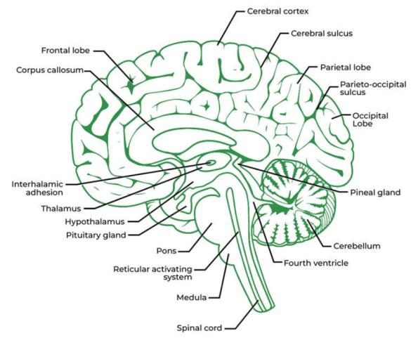
- Forebrain: It is divided into three sections, which are as follows:
- Olfactory lobes: These are a pair of very tiny, solid club-shaped entities that are far apart from one another
- Cerebrum – It is the largest and most complex. The cerebral hemispheres on the right and left of the brain are separated by a deep gap and are joined by the corpus callosum, a network of myelinated fibers.Diencephalon
- It encloses the third ventricle, which is a slit-like chamber. The thick right and left sides of this cavity are referred to as the thalami, its thin floor as the hypothalamus, and its thin ceiling as the epithalamus.
- Midbrain: It is placed between the thalamus/hypothalamus of the forebrain and the pons of the hindbrain. Its upper surface has two pairs of spherical extensions called corpora quadrigemina and two bundles of fibers termed crura cerebri.
- Hindbrain: It is made up of:
- Cerebellum: The cerebellum is the second largest portion of the human brain. It is made up of two lateral cerebellar hemispheres and a central worm-shaped component called the vermis. The cerebellum has grey matter on the outside, which is composed of three layers of cells and fibers. It also has Golgi cells, basket cells, and granule cells.
- Pons Varolii: The pons Varolii is an oval mass that lies above the medulla oblongata. It is mostly made up of nerve fibers that connect various areas of the brain.
- Medulla oblongata: This structure extends from the pons Varolii above and connects to the spinal cord below. The midbrain, pons Varolii and medulla oblongata are referred to as the brain stem.
For More Information Read: Human Brain
Q 2: Compare the following:
- (a) Central neural system (CNS) and Peripheral neural system (PNS)
- (b) Resting potential and action potential
Answer:
Comparision between Central neural system (CNS) and Peripheral neural system (PNS) is given below:
| Aspect |
Central Nervous System (CNS) |
Peripheral Nervous System (PNS) |
| Location |
Brain and Spinal Cord |
Nerves outside the brain and spinal cord |
| Function |
Processes and integrates information, initiates and coordinates responses |
Transmits sensory information to the CNS and carries motor commands from the CNS to muscles and glands |
| Components |
Brain and Spinal Cord |
Cranial nerves and spinal nerves |
| Protection |
Protected by bones (skull and vertebrae), cerebrospinal fluid, and meninges |
Lacks bony protection; some protection provided by connective tissue coverings |
| Types of Cells |
Neurons and Glial Cells |
Mostly neurons |
| Control of Functions |
Controls voluntary and involuntary actions, thoughts, and emotions |
Primarily involved in involuntary actions and reflexes |
| Neurotransmitter Activity |
Synaptic transmission |
Synaptic transmission |
| Response Time |
Generally slower response time compared to the PNS |
Generally faster response time compared to the CNS |
Comparision between Resting potential and action potential is given below:
| Aspect |
Resting Potential |
Action Potential |
| Definition |
Stable charge of a neuron at rest |
Brief reversal of membrane potential |
| Membrane Potential |
Around -70 mV |
Rapid depolarization to around +40 mV |
| Ion Movement |
Maintained by ion gradients |
Rapid influx of Na+ and efflux of K+ |
| Channels Involved |
Mostly leak channels and pumps |
Voltage-gated Na+ and K+ channels |
| Stimulus |
Spontaneous occurrence |
Threshold stimulus required |
| Duration |
Persistent |
Short-lived |
| Direction of Ion Movement |
Minimal movement |
Rapid movement |
| Role in Neuronal Signaling |
Establishes baseline |
Mediates electrical signaling |
Q3: Explain the following processes:
- (a) Polarisation of the membrane of a nerve fiber.
- (b) Depolarisation of the membrane of a nerve fiber.
- (c) Transmission of a nerve impulse across a chemical synapse.
Solution:
- (a) Polarisation of a nerve fiber membrane:
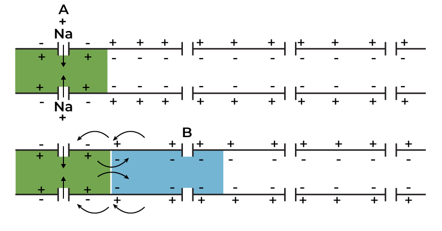
When a nerve fiber is at rest, it is considered to be in a polarised state. The nerve fiber’s membrane has a resting potential when it is polarised. The following steps occur during the polarization of a nerve fiber’s membrane:
- When a depolarised area of a nerve fiber first begins to become polarised, there are more K + ions outside the nerve fiber and a considerable amount of Na + ions in the axon membrane.
- As the membrane region gets polarised, it becomes more permeable to K + ions and impenetrable to Na + ions and negatively charged proteins.
- A sodium-potassium pump sends 3 Na + ions outside the axon and 2 K + ions into the axon via active transport.
- Because of the flow of sodium and potassium ions, the inner side of the membrane becomes electronegative (negatively charged), while the outer side becomes electropositive (positively charged). This causes the nerve fiber to become polarised.
Also Read: Generation And Conduction Of Nerve Impulse
- (b) Depolarisation of the membrane of a nerve fiber.
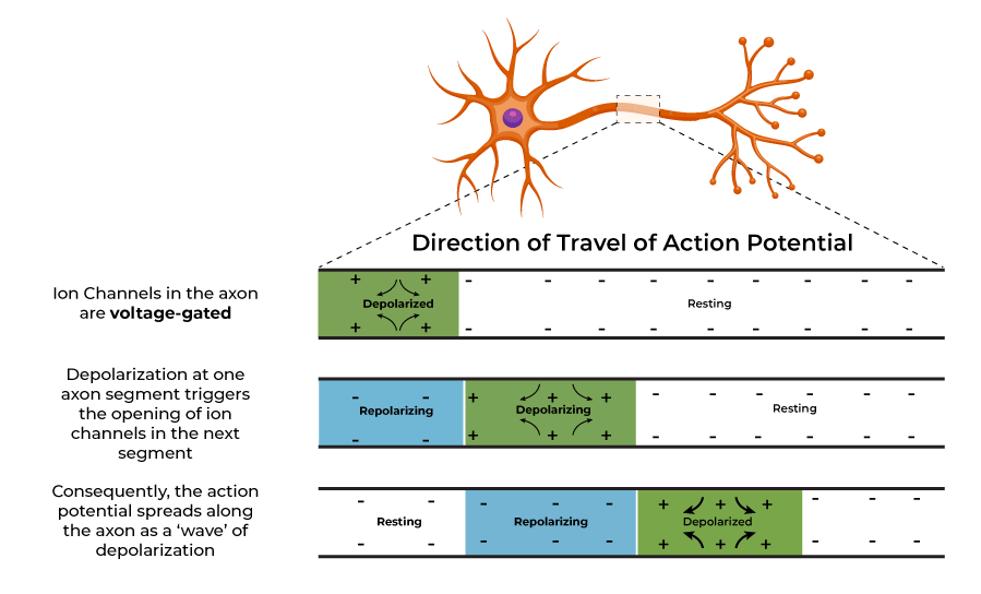
When a nerve fiber is activated, it is said to be in a depolarised state. The membrane of the nerve fiber undergoes an action potential when it is depolarized.
During the process of depolarisation of a nerve fiber’s membrane, the following phases occur:
- The axon has a higher concentration of K + ions in a polarised condition, while the concentration of Na + ions is higher outside the axon.
- When a nerve fiber is stimulated, the permeability of the membrane for Na + ions and K + ions reverses.
- The membrane becomes highly permeable to Na + ions.
- There is a fast influx of Na + ions into the axon.
- As a result, the inside of the membrane becomes positively charged, while the exterior of the membrane gets negatively charged.
- Finally, the nerve fiber’s membrane depolarizes and it experiences an action potential.
(c) Transmission of a nerve impulse across a chemical synapse.
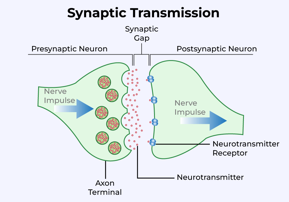
- The membranes of the pre-synaptic and post-synaptic neurons combine to form a synapse.
- A gap known as the synaptic cleft may or may not divide two synapses.
- The synaptic cleft separates the pre-and post-synaptic neurons at a chemical synapse.
- The calcium ions in the synaptic cleft enter the synaptic knobs at the axon terminals of the pre-synaptic neuron when an impulse reaches the axon terminal.
- The synaptic vesicles in the pre-synaptic neuron’s synaptic knobs migrate in its direction and fuse with the plasma membrane.
- Acetylcholine, a neurotransmitter, is released by the vesicles in the synaptic cleft. (Empty synaptic vesicles are filled when they return to the cytoplasm of the pre-synaptic neuron.)
- The protein receptors found on the plasma membrane of post-synaptic neurons are where the molecules of acetylcholine bind.
- As a result of this interaction, potassium ions exit the post-synaptic membrane and sodium ions enter the post-synaptic neuron.
- This causes the membrane of the post-synaptic neuron to create an action potential, which then transmits the impulse to the post-synaptic neuron.
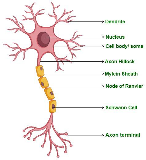
Q4: Draw labeled diagrams of the following:
Answer:
(a) The labelled diagram of Neuron is given below:
(b) Brain
Q5: Write short notes on the following:
- (a) Neural coordination
- (b) Forebrain
- (c) Midbrain
- (d) Hindbrain
- (e) Synapse
Answer:
(a) Neural Coordination
Neurons are highly specialized cells that provide neural synchronization. The neurological system functions by nerve impulses and is made up of a network of point-to-point connections between the neurons and the organs. The input and the response—receptors, and effectors—are always coordinated by the neural system. Neural coordination carries out and manages every bodily function. The skin, among other organs, receives the stimulation, and a reaction is produced and conveyed to the muscles or glands. The brain system constantly stores the prior stimulus in memory. Running, walking, writing, and talking are examples of voluntary actions that benefit from neural coordination’s control and symmetry.
(b) Forebrain
The cerebrum, thalamus, and hypothalamus are three components of the cortex.
- Cerebrum: It is the major component of the brain. A fissure in the cerebrum divides the left and right cerebral hemispheres. The corpus callosum connects the hemispheres of the brain. The cerebral cortex is a layer of visible folds made up of cells that cover the cerebral hemisphere. Due to its greyish coloring, it is referred to as grey matter. Many areas of the cerebral cortex don’t appear to have any sensory or motor functions. Association areas carry out a number of complex tasks, such as memory, communication, and intercessory associations. The fibers of the tract are shielded by the myelin coating in the center of the cerebral hemisphere. Because of its impenetrable aspect, white matter is so named. The association areas are in charge of memory, communication, and intercessional associations.
- Thalamus: The Thalamus is a section of the cerebrum that wraps around the center of the forebrain. At this center, sensory and motor signaling are synchronized.
- Hypothalamus: The hypothalamus contains a number of structures that control body temperature, hunger, and thirst. It is linked to the pituitary gland and regulates both growth and sexual behaviors.
Also Read: Difference Between Cerebellum And Cerebrum
(c) Midbrain
It is positioned between the pons of the hindbrain and the thalamus/hypothalamus of the forebrain The midbrain is connected to the brain by a passageway known as the cerebral aqueduct. The corpora quadrigemina, four circular lobes that make up the majority of the dorsal section of the midbrain, are present. The brain stem is made up of the hindbrain and midbrain.
(d) Hindbrain
Pons, cerebellum, and medulla make up the hindbrain. Pons is made up of fiber lines that link various brain areas. In order to accommodate more neurons, the surface of the cerebellum is extremely twisted. The spinal cord and the brain’s medulla are interconnected. Respiratory, cardiovascular, and gastric secretion control centers are located in the medulla.
(e) Synapse
The membranes of the pre-synaptic and post-synaptic neurons combine to form a synapse. A gap known as the synaptic cleft may or may not divide two synapses. Electrical and chemical synapses are two different types of synapses.
Q6: Give a brief account of the Mechanism of Synaptic Transmission.
Answer:
A synapse is the junction of two neurons. It is separated by a cleft and exists between the dendrite of one neuron and the axon terminal of the following neuron. There are two ways synaptic transmission happens.
- Chemical Transmission: When a nerve impulse reaches the endplate of an axon, acetylcholine, a neurotransmitter, is released across the synaptic cleft. This substance is created in the cell body of the neuron and sent to the axon terminal. Over the cleft, acetylcholine diffuses and binds to receptors on the membrane of the following cell. Depolarization of the membrane and the beginning of an action potential follow from this.
- Electrical Transmission: In this type of transmission, an electric current is generated in the neuron. The action potential that is brought on by this electric current results in the transmission of nerve impulses across the nerve fiber. This form of nerve conduction is more rapid than the chemical process.
Q7: Explain the Role of Na+ in the generation of the action potential.
Answer:
Due to the electrochemical gradient, sodium ions diffuse into the intracellular fluid from the outside. The membrane changes from being positively charged on the inside to being negatively charged outside as the potassium ions exit. The membrane is said to be depolarized as a result of the action potential, a quick shift in the membrane potential.
Read Also: Membrane potential
Q8: Differentiate between:
- (a) Myelinated and non-myelinated axons
- (b) Dendrites and axons
- (c) Thalamus and Hypothalamus
- (d) Cerebrum and Cerebellum
Answer:
(a) Myelinated and non-myelinated axons vs Non-myelinated Axons are:
| Aspect |
Myelinated Axons |
Non-myelinated Axons |
| Structure |
Covered with myelin sheath, speeding up signal transmission |
Lack myelin sheath, slowing down signal transmission |
| Conduction Speed |
Faster due to “jumping” action potentials between nodes |
Slower as action potentials propagate along the entire axon |
| Energy Efficiency |
More efficient with less ion leakage through myelin |
Less efficient with more ion leakage |
| Protection |
Better insulation and protection against damage |
More vulnerable to damage and signal loss |
| Examples |
Found in central and peripheral nervous system |
Found in sensory neurons, especially for pain perception |
- (b) Dendrites and axons vs axons are give below:
| Aspect |
Dendrites |
Axons |
| Function |
Receive incoming signals from other neurons |
Transmit signals away from the cell body to other neurons or cells |
| Structure |
Branched extensions from the cell body |
Single, elongated projection from the cell body |
| Size |
Typically shorter and more numerous |
Longer, with variable lengths |
| Direction of Signal Flow |
Signal reception towards the cell body |
Signal transmission away from the cell body |
| Presence of Myelin |
Usually non-myelinated |
Can be myelinated or non-myelinated |
| Synaptic Terminals |
Rarely have synaptic terminals |
Have synaptic terminals at the axon terminals for signal transmission |
| Role in Neuronal Signaling |
Integrates and processes incoming signals |
Transmits signals to other neurons or effector cells |
- (c) Thalamus vs Hypothalamus is given below:
| Aspect |
Thalamus |
Hypothalamus |
| Location |
Situated above the brainstem |
Located below the thalamus |
| Function |
Acts as a relay station for sensory information, directing it to the appropriate areas of the cerebral cortex |
Regulates homeostasis, hormone production, autonomic nervous system, and emotional responses |
| Hormonal Control |
Not primarily involved in hormonal regulation |
Controls hormone secretion through the pituitary gland |
| Role in Sleep/Wake Cycle |
Helps regulate sleep and wakefulness indirectly |
Directly regulates sleep and wakefulness through the suprachiasmatic nucleus |
| Relationship with Pituitary |
No direct connection with the pituitary gland |
Directly connected to the pituitary gland via the hypothalamic-pituitary axis |
- (d) Cerebrum vs Cerebellum is as follows:
| Aspect |
Cerebrum |
Cerebellum |
| Location |
Largest part of the brain, located in the uppermost region |
Located below the cerebrum, at the back of the brain |
| Function |
Responsible for higher brain functions including cognition, sensory perception, and voluntary motor control |
Coordinates voluntary movements, balance, posture, and motor learning |
| Structure |
Comprises two hemispheres connected by the corpus callosum |
Consists of two hemispheres with a highly folded surface called the cerebellar cortex |
| Gray Matter |
Contains the cerebral cortex, responsible for conscious thoughts and information processing |
Contains the cerebellar cortex, involved in integrating sensory and motor information |
| White Matter |
Comprises nerve fibers connecting different regions of the brain |
Comprises nerve fibers connecting the cerebellum to other parts of the brain and spinal cord |
| Motor Control |
Involved in initiating and coordinating voluntary muscle movements |
Fine-tunes and regulates motor movements initiated by the cerebrum |
| Cognitive Functions |
Essential for language, memory, attention, and decision-making |
Supports procedural memory and motor coordination, but less involved in cognitive functions |
Q9: Answer the following:
- (a) Which part of the human brain is the most developed?
- (b) Which part of our central neural system acts as a master clock?
Answer:
- (a)The most developed part is the cerebrum.
- (b)The central nervous system’s hypothalamus serves as a master clock.
Q10: Distinguish between
- (a) afferent neurons and efferent neurons
- (b) impulse conduction in a myelinated nerve fiber and unmyelinated nerve fiber
- (c) cranial nerves and spinal nerves.
Answer:
- (a) The difference between Afferent neurons and Efferent neurons are:
| Aspect |
Afferent Neurons |
Efferent Neurons |
| Direction of Signal Flow |
Carry sensory information to the CNS |
Transmit signals from CNS to muscles/glands |
| Function |
Sense external stimuli |
Control movement and glandular secretion |
| Examples |
Skin, eyes, ears sensory neurons |
Motor neurons for muscles and glands |
| Pathway |
Signal travels to CNS |
Signal originates from CNS |
| Synaptic Connections |
Connect with interneurons in CNS |
Directly connect with target cells |
- (b) The difference between impulse conduction in a myelinated nerve fiber and unmyelinated nerve fiber:
| Aspect |
Myelinated Nerve Fiber |
Unmyelinated Nerve Fiber |
| Conduction Speed |
Faster due to saltatory conduction, where impulses “jump” between nodes of Ranvier |
Slower as impulses propagate continuously along the entire length of the fiber |
| Energy Efficiency |
More energy-efficient with reduced ion leakage through myelin sheath |
Less energy-efficient with more ion leakage along the fiber |
| Protection |
Provides insulation and protection, minimizing signal loss and damage |
Lacks insulation and is more susceptible to signal loss and damage |
| Examples |
Found in motor neurons and sensory neurons responsible for rapid responses |
Found in pain receptors and sensory neurons requiring continuous signal transmission |
- (c) The difference between cranial nerves and spinal nerves are:
| Aspect |
Cranial Nerves |
Spinal Nerves |
| Location |
Arise from the brain and exit the skull through openings called foramina |
Emerge from the spinal cord and exit the vertebral column through spaces between vertebrae called intervertebral foramina |
| Number |
12 pairs |
31 pairs (8 cervical, 12 thoracic, 5 lumbar, 5 sacral, 1 coccygeal) |
| Function |
Primarily innervate structures of the head and neck, controlling sensory and motor functions |
Innervate the rest of the body, controlling both sensory and motor functions |
| Composition |
May contain sensory, motor, or mixed (both sensory and motor) fibers |
Typically mixed nerves containing both sensory and motor fibers |
| Examples |
Olfactory nerve (I), optic nerve (II), facial nerve (VII) |
Sciatic nerve, median nerve, ulnar nerve |
Related Links
FAQs on NCERT Solutions for Class 11 Chapter 18 – Neural Control and Coordination Class 11
Where Can I Find the Neural Control and Coordination Class 11 Solutions?
The solutions for chapter 18 Neural Control and Coordination is given at the top of this article.
What are the Advantages of using NCERT Solutions for Class 11 Biology Chapter 18?
The advantages of using NCERT solutions for chapter 18 is that it provides comprehensive coverage of the chapter’s topics and offers accurate explanations and solutions suited to the NCERT curriculum.
What are the Important Topics of NCERT Solutions for Class 11 Biology Chapter 18?
Chapter 18 of NCERT Solutions for Class 11 Biology highlights essential topics including the Neural System, Human Neural System, Neuron as the Structural and Functional Unit, Central Neural System, Reflex Action, Reflex Arc, and Sensory Reception and Processing, pivotal for board exam preparation.
Where to Class 11 Neural Control and Coordination NCERT PDF?
You can find the pdf of Neural Control and Coordination chapter 18 here.
Share your thoughts in the comments
Please Login to comment...