NCERT Solutions for Class 11 Biology Chapter 7 Structural Organisation in Animals
Last Updated :
19 May, 2023
NCERT Solutions for class 11 Chapter 7 Structural Organisation in Animals: The chapter on Structural Organisation in Animals is important for students approaching the home exams. This article introduces NCERT solutions designed to help students explain the concepts of further learning and how to write to get good grades on exams. The solutions are presented in very simple language for ease of understanding.
NCERT CBSE Chapter 07 Structural Organisation In Animals of Class 11 explains that the life processes of the human body are functional at various levels of structural organization which include cellular, tissue, organ, organ system, and the organism level. Higher organizations are built from the lower levels of the organization. The study of the structures and their organization in a body is known as Anatomy. This is associated with a proper understanding of the structures based on the formation of specific systems like digestive, circulatory, excretory, etc. Revise the basic concepts of Structural Organisation in Animals for quick revision and class notes.
Questions of Chapter 7 Structural Organisation in Animals (Based on the New Syllabus)
Q1: Draw a neat diagram of the Digestive System of a Frog.
Answer:
The digestive system of a frog is shown below:

Q2: Mention the Function of the Ureters in Frogs.
Answer:
Ureters in frogs: They help in sperm and urine transport.
Questions of Chapter 7: Structural Organisation in Animals (Based on the Old Syllabus)
Q1: Answer in One Word or One Line.
- Give the common name of Periplanata americana.
- How many spermathecae are found in earthworms?
- What is the position of ovaries in cockroaches?
- How many segments are present in the abdomen of cockroaches?
- Where do you find Malpighian tubules?
Answer:
- Cockroach
- Four pairs
- In 2nd to 6th abdominal segments. Ovaries are two in number and are located laterally.
- 10 segments
- Malpighian tubules are the excretory organs, present at the junction of the midgut and hindgut of the alimentary canal.
Q2: Answer the Following.
- What is the Function of Nephridia?
- How many types of Nephridia are found in Earthworms Based on their Location?
Answer:
- (i) Nephridia maintains Osmoregulation and helps to excrete out the waste products in many invertebrates.
- (ii) There are three types of Nephridia found in the Earthworms are:
- Septal nephridia: They are situated on the inter-segmental septum between the 15th and 16th segments.
- Integumentary nephridia: They are situated in all the segments except the first two segments.
- Pharyngeal nephridia: They are situated as a cluster of three pairs in the 4th, 5th, and 6th segments.
Q3: Draw a Labeled Diagram of the Reproductive Organs of an Earthworm.
Answer:
A well-labeled diagram of the reproductive organs of an earthworm is:
 Reproductive organs of an earthworm
Q4: Draw a Labeled Diagram of the Alimentary Canal of a Cockroach.
Answer:
A well-labeled diagram of the alimentary canal of a cockroach is:
 The alimentary canal of a cockroach
Q5: Distinguish Between the Following:
- (a) Prostomium and Peristomium
- (b) Septal nephridium and Pharyngeal nephridium
Answer:
(a) Difference Between Prostomium and Peristomium
|
Prostomium
|
Peristomium
|
|
A lobe arises from the peristomium
|
This is the very first segment
|
|
It projects the mouth
|
The mouth is located on the ventral side of the first segment
|
(b) Diffrence Between Septal nephridium and Pharyngeal nephridium
|
Septal nephridium
|
Pharyngeal nephridium
|
|
These are present on both the sides of the intersegment septa from the 15th septa to the last.
|
These are present as the clusters in 4th, 5th, and 6th segments.
|
|
They open into the intestine
|
They open into the the pharynx
|
Q6: What are the Cellular Components of Blood?
Answer:
The cellular components of blood are:
- Reb blood cells or erythrocytes: They transport oxygen from the lungs to the other body parts.
- White blood cells or leukocytes: They are known as the soldiers of our body because they fight against infections and assist in the immune process.
- Platelets or thrombocytes: They help in blood clotting at the time of injury.
Q7: What are the Following, and where do you find them in an Animal Body?
- (a) Chondrocytes
- (b) Axons
- (c) Ciliated epithelium
Answer:
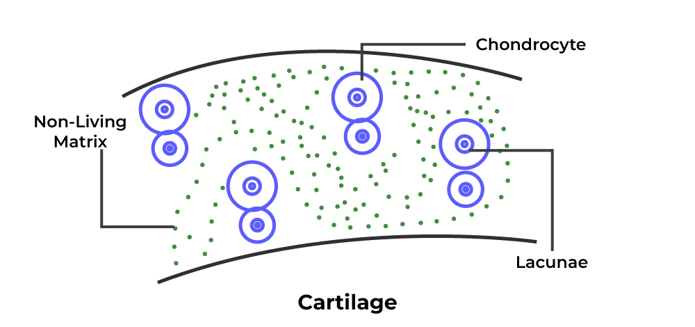
(a) Chondrocytes: Cartilage is formed by some special cells known as Chondrocytes. Cartilage is a strong and flexible connective tissue that provides protection to the joints and bones by acting as a shock absorber. Location– They are located in the matrix or ground base of the cartilage.
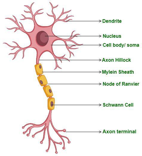
(b) Axons– An axon or nerve fiber is the elongated part of a nerve cell or neuron that carries nerve impulses or signals away from the cell body. A typical neuron has an axon that connects it with other neurons or muscles or glands. Location– Axons are located between the cell body and axon terminals or ends.
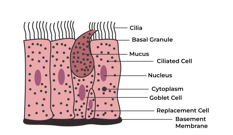
(c) Ciliated epithelium– Ciliated Epithelium is a thin tissue that has some hair-like structure known as cilia, which helps in the locomotion of our body. Location– They are located in the human respiratory tract and the fallopian tubes of women.
Q8: Describe Various Types of Epithelial Tissues with the Help of Labeled Diagrams.
Answer:
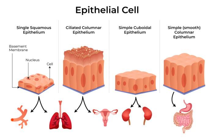
The epithelium is a type of body tissue that lines the covering on all internal and external surfaces of your body which includes body cavities and hollow organs. Epithelial tissues are formed of a compact layer of cells. One surface of the epithelial tissue is attached to the external environment or body fluid. The other surface is attached to the tissue. There are two types of epithelial tissues:
- Simple epithelial tissues: These consist of a single layer of cells resting on the basement membrane. It lines the body cavity, ducts, and tubes.
- Compound epithelial tissues: Compound epithelium tissue is shaped of numerous layers of epithelial cells of dissimilar shapes representing newly shaped and mature cells. This tissue forms the covering of all body surfaces and they are the major tissue in glands.
Simple epithelial tissues are divided into the following types:
- Columnar epithelium: It is made up of cells that appear like columns that line the small intestine and ducts of many glands.
- Cuboidal epithelium: It is made up of cells that are cube-like in appearance and line the kidney tubules
- Squamous epithelium: It is made up of thin sheets-like or flat cells which give protection against microorganisms. The outer layer of the skin (the epidermis) is made of stratified squamous epithelial cells. The lining of the blood vessels is also made up of these tissues.
- Ciliated Epithelium: They are made up of rectangular cells which line the respiratory tract.
Q9: Distinguish Between
- (a) Simple epithelium and compound epithelium
- (b) Cardiac muscle and striated muscle
- (c) Dense regular and dense irregular connective tissues
- (d) Adipose and blood tissue
- (e) Simple gland and compound gland
Answer:
(a) Difference Between Simple Epithelium and Compound Epithelium
| Simple Epithelium |
Compound Epithelium |
|
It is made up of single-layered cells.
|
It is made up of multilayered cells.
|
|
They include squamous, cuboidal, columnar, and ciliated epithelium.
|
They include stratified and transitional epithelium.
|
(b) Difference Between Cardiac Muscle and Striated Muscle
| Cardiac muscle |
Striated muscle |
|
They are present in the wall of the heart.
|
They are present in the tongue, pharynx, limbs, etc.
|
|
They are made up of unbranched fibers.
|
They are made up of branched fibers.
|
(c) Different Between Dense Regular and Dense Irregular Connective Tissues
|
Dense regular connective tissues
|
Dense irregular connective tissues
|
|
They are made up of regularly oriented fibers.
|
They are made up of irregularly oriented fibers.
|
|
They are present in tendons, ligaments, etc.
|
They are present in the dermis of the skin.
|
(d) Different Between Adipose and Blood Tissue
|
Adipose tissue
|
Blood tissue
|
|
It is present below the skin, kidney, and in the bone marrow.
|
It is present in the blood.
|
|
It stores fat and helps in the maintenance of body heat.
|
It helps in the transportation of oxygen, food components, etc.
|
(e) Different Between Simple Gland and Compound Gland
|
Simple gland
|
Compound gland
|
|
It is unicellular, i.e. made up of one cell, or multicellular, i.e. made up of many cells
|
It is multicellular.
|
|
It may be branched or unbranched. Examples are sweat gland, sebaceous gland, etc.
|
It has branches and sub-branches like primary, secondary, and tertiary. An example is mammary glands
|
Q10: Mark the odd one in each series.
- (a) Areolar tissue; blood; neuron; tendon
- (b) RBC; WBC; platelets; cartilage
- (c) Exocrine; endocrine; salivary gland; ligament
- (d) Maxilla; mandible; labrum; antennae
- (e) Protonema; mesothorax; metathorax; coxa
Answer:
- (a) Neuron: It is not a connective tissue. It is a nervous tissue.
- (b) Cartilage: It is not a component of the blood while the rest are the components of the blood.
- (c) Ligament: It is a connective tissue while the rest are the glands.
- (d) Antennae: They are associated with sensory functions while the rest are the mouth parts, involved in digestive functions.
- (e) Protonema: It is a part of the life cycle in mosses while the rest parts are the leg segments in cockroaches.
Q11: Match the terms in column I with those in column II.
Column I
|
Column II
|
(a) Compound epithelium
|
(i) Alimentary canal
|
(b) Compound eye
|
(ii) Cockroach
|
(c) Septal nephridia
|
(iii) Skin
|
(d) Open circulatory system
|
(iv) Mosaic vision
|
(e) Typhlosole
|
(v) Earthworm
|
(f) Osteocytes
|
(vi) Phallomere
|
(g) Genitalia
|
(vii) Bone
|
Answer:
| Column I |
Column II |
|
(a) Compound epithelium
|
(i) Skin
|
|
(b) Compound eye
|
(ii) Mosaic vision
|
|
(c) Septal nephridia
|
(iii) Earthworm
|
|
(d) Open circulatory system
|
(iv) Cockroach
|
|
(e) Typhlosole
|
(v) Alimentary canal
|
|
(f) Osteocytes
|
(vi) Bone
|
|
(g) Genitalia
|
(vii) Phallomere
|
Q12: Mention the Circulatory System of Earthworms Briefly.
Answer:
- The circulatory system of earthworms is of a closed type i.e. blood flows in the blood vessels and capillaries.
- Blood consists of blood plasma and blood cells.
- Haemoglobin is present in the blood which gives red colour to the blood.
- Blood circulation occurs in one direction only i.e. unidirectional.
- On the 4th, 5th, and 6th segments, blood glands are present. which produces blood cells, hemoglobin which is dissolved in the blood plasma.
Q13: Draw a Neat Diagram of the Digestive System of a Frog.
Answer:
The diagram of the digestive system of a frog is:
Q14: Mention the Function of the following (a) Ureters in Frogs (b) Malpighian Tubules (c) Body Wall in Earthworms.
Answer:
- (a) Ureters in frogs: They help in sperm and urine transport.
- (b) Malpighian tubules: They help in osmoregulation (reabsorption of water, ions, and solutes) and production of urine.
- (c) Body wall in earthworms: It helps in burrowing and bodily movements.
Share your thoughts in the comments
Please Login to comment...