Mechanism of Muscle Contraction – CBSE Class 11
Last Updated :
07 Aug, 2023
The mechanism of muscle contraction refers to the process by which muscles generate force and produce movement. Muscle contraction involves a complex interplay of biochemical and physiological events within the muscle fibers. When a nerve signal reaches a muscle, it triggers the release of calcium ions, which allows actin and myosin proteins to interact and causes these filaments to slide past each other. This sliding filament mechanism shortens the muscle fibers and results in contraction and the generation of force.
Sarcomere Structure
The sarcomere is the basic functional unit of muscle contraction. It is located between two Z-lines and consists of thin and thick myofilaments. The arrangement of these filaments within the sarcomere is responsible for the characteristic striated appearance of skeletal muscle fiber. In a Sarcomere, each thin myofilament is surrounded by 3 thick myofilaments and each thick myofilament is surrounded by 6 thin myofilaments. Sarcomere is divided into certain bands and zones based on the presence or absence of thick or thin filaments:
- A-band: A-Band is also known as an anisotropic or dark band because light does not pass through it. It is the region where the thick filament is present.
- H-Zone: H-Zone is also known as Henson’s Zone. It is the non-overlapping region between the A-Band. It is located in the middle of the sarcomere where only thick filament is present. It can also be defined as the central gap between actin filaments extending through myosin filaments in A-Band.
- O-Zone: O-Zone is the region where both actin and myosin are present. It is also present within A-Band.
- I-Band: I-Band is also known as an isotropic band or light band. It is the region where only thin filament is present. It is part of two sarcomeres. It is divided into two halves by the Z- line.
In a Sarcomere, each thin myofilament is surrounded by 3 thick myofilaments and each thick myofilament is surrounded by 6 thin myofilaments.

Sliding Filament Theory
The sliding filament theory is given by H.F. Huxley and A.F. Huxley. It provides an explanation for how sarcomere contracts. According to this theory, filaments do not contract. Muscle contraction is the result of the thin filaments sliding past the thick filaments. Thick filaments do not move.
Changes in Sarcomere during contraction:
- Z-lines come closer.
- H-Zone decreases
- I-Band decreases
- O-Zone increases
- The A-Band remains the same.
Mechanism of Muscle Contraction
Muscle contraction involves a series of steps that allow the actin and myosin filaments to slide past each other which results in the shortening of the sarcomere. Following are the steps involved in muscle contraction:
Excitation
The process of muscle contraction begins with excitation, which involves the initiation of an electrical impulse called an action potential. This action potential originates from a motor neuron in the brain or spinal cord and travels until it reaches the neuromuscular junction, also known as the motor end plate. It is the region where the motor neuron connects with the muscle fiber.
Neuromuscular Junction
At the neuromuscular junction, the action potential triggers the release of a neurotransmitter called acetylcholine into the synaptic cleft. Acetylcholine binds to receptors on the sarcolemma, which leads to the generation of another electrical impulse along the sarcolemma.
Transmission by T-tubules
The electrical impulse propagates along the sarcolemma and goes throughout the muscle cell through invaginations in sarcolemma called T-tubules. These T-tubules allow the electrical impulse to reach the interior of the muscle fiber and ensure simultaneous activation of the whole muscle cell.
Release of Calcium Ions
The transmembrane signal in the T-tubules results in the release of calcium ions from the sarcoplasmic reticulum. The calcium ions diffuse into the cytoplasm
Calcium-Binding to Troponin
Within the muscle fiber, the calcium ions bind to a regulatory protein called troponin present in thin filaments. This binding causes a conformational change and exposes the myosin-binding sites on the actin filaments making it ready for contraction.
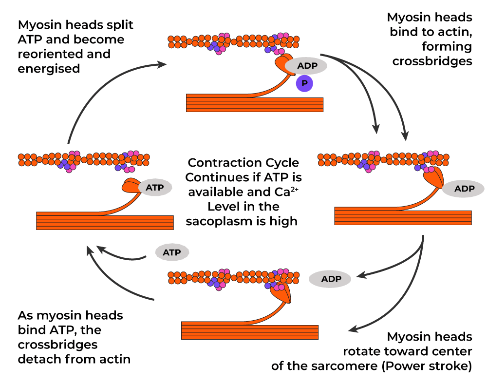
Cross-Bridge Formation
ATP is hydrolyzed into ADP and inorganic phosphate in the presence of Mg2+ and Ca2+ ions. When ADP is removed, the head of meromyosin binds strongly with myosin binding sites on the actin filaments. This is known as Cross-Bridge Formation or Actomyosin interaction or Power Stroke.
Sliding of Filaments
When inorganic phosphate is removed, the head of the meromyosin moves backward and it pulls thin filaments inside, towards the center of the sarcomere. Thin filaments slide over thick filaments and thick filaments do not slide. This sliding movement shortens the sarcomeres.
Detachment of Cross-Bridge
After the power stroke, ATP is formed using high-energy phosphate like phosphocreatine. In this process, Creatinine (excretory product) is formed as a by-product. This ATP binds with the ATP binding site on the head of meromyosin. This results in the detachment of actin and myosin.
These steps like attachment, power stroke, detachment, and reactivation of myosin heads repeat and this cycle continues as long as the calcium concentration remains high and ATP is available.
Muscle Relaxation
When Relaxation is required, the stimulation from the motor neuron stops and Acetylcholine, already present in the cleft, is digested by the Acetylcholinesterase enzyme. Also, the calcium ions are actively pumped back into the sarcoplasmic reticulum using the Calsequestrin pump, which reduces their concentration in the cytoplasm. As a result, inhibitor troponin (TpI) rolls backward and tropomyosin again covers the myosin-binding sites on the actin filaments. This prevents further cross-bridge formation and stops muscle contraction.
Conclusion
Muscle contraction is a complex and highly coordinated process that enables us to move and perform daily activities. Understanding the mechanism of muscle contraction, including the sarcomere structure, the process of contraction, and the sliding filament theory, provides an understanding of the processes of locomotion and movement in our bodies.
FAQs on Mechanism of Muscle Contraction
Q: What initiates muscle contraction?
Answer:
The impulse originates from a motor neuron and travels to the neuromuscular junction initiates muscle contraction.
Q: How does the release of calcium ions contribute to muscle contraction?
Answer:
The calcium ions released from sarcoplasmic reticulum bind to troponin and cause a conformational change that exposes the myosin-binding sites on actin filaments.
Q: What is the role of ATP in muscle contraction?
Answer:
ATP is essential for muscle contraction. It is hydrolyzed into ADP, inorganic phosphate and release energy that helps in the movement of myosin heads.
Q: What is the power stroke in muscle contraction?
Answer:
ATP is hydrolyzed into ADP and inorganic phosphate in the presence of Mg2+ and Ca2+ ions. When ADP is removed, the head of meromyosin binds strongly with myosin binding sites on the actin filaments. This is known as Power Stroke.
Share your thoughts in the comments
Please Login to comment...